Contents
Scroll to:
https://doi.org/10.37489/2588-0527-0008
EDN: BQRZEP
Scroll to:
Background. In the context of predictive medicine, personalized assessment of ischemic stroke risk based on molecular genetic testing is becoming increasingly relevant. Polymorphisms in folate cycle genes (MTHFR, MTR, MTRR), leading to hyperhomocysteinemia, cause endothelial dysfunction and accelerate atherogenesis. The role of the renin-angiotensin system (RAS) is no less important: genetic variations in the AGT and NOS3 genes are associated with arterial hypertension and altered vascular tone, which creates a predisposition to cerebrovascular accidents.
Objective. To identify associations between polymorphisms of folate cycle and renin-angiotensin system genes and the development of acute ischemic stroke and transient ischemic attack (TIA).
Methods. The observational study included 492 patients who were treated in hospital at the Institution Clinical Hospital No. 1 of the Presidential Administration of the Russian Federation in the period 2022–2025 and who signed informed consent for molecular genetic testing. A comparative analysis of clinical, laboratory, instrumental, and molecular genetic data (polymorphisms of MTHFR, MTR, MTRR, AGT, NOS3, and AGTR1) was performed between the ischemic stroke group (n=426) and the TIA group (n=66). Statistical analysis was carried out using the Mann-Whitney U test and Pearson's chisquared test.
Results. Stroke patients were older (73 vs 66 years, p=0.005) and more frequently male (43.4 % vs 27.3 %, p=0.013). The stroke group had significantly higher triglyceride levels (p < 0.001) and potassium levels (p < 0.001), as well as a higher frequency of pathological MRT findings (p < 0.001). Genetic analysis revealed a significant predominance in the stroke group of homozygous CC genotypes for MTHFR C677T (p=0.001), AGT174 (p=0.004), AGT235 (p < 0.001), and NOS3 (p=0.028). In contrast, the TIA group more frequently carried TT genotypes for C677T AGT235, and NOS3, as well as the heterozygous CT genotype for AGT174.
Conclusion. Molecular genetic testing of polymorphisms in folate cycle and renin — angiotensin system genes represents a promising tool for risk stratification and differential diagnosis of ischemic stroke versus TIA, and may serve as a foundation for personalized pharmacotherapy in the nearest future.
Kovtun N.A., Isaeva Т.V., Savelyeva M.I., Boyarintsev V.V. The role of single nucleotide polymorphisms in predicting individual stroke risk. Pharmacogenetics and Pharmacogenomics. 2026;(2):24-34. (In Russ.) https://doi.org/10.37489/2588-0527-0008. EDN: BQRZEP
Despite advances in modern neurology in the diagnosis and treatment of acute cerebrovascular accidents, the prognosis of the disease and the likelihood of its development in a particular patient remain largely individualized. One of the key factors in such variability is hereditary characteristics that determine predisposition to thrombosis and vascular complications [1].
A key link in the pathogenesis of ischemic stroke (IS) is thrombotic occlusion of a cerebral vessel, which makes genes of the hemostasis system of particular interest. However, the role of hereditary thrombophilia is not limited to venous thrombosis but also extends to the arterial bed [2]. Polymorphisms of coagulation factor genes (F2, F5), platelet receptor genes (ITGA2, ITGB3), and plasminogen activator inhibitor-1 (PAI-1) genes can alter the coagulation potential of blood, increasing the tendency to thrombus formation or, conversely, to hemorrhagic complications [3].
At the same time, genetically determined metabolic disorders make a significant contribution to the development of vascular pathology. The condition of the vascular wall, which serves as the substrate for atherothrombosis, directly depends on biochemical homeostasis. Polymorphisms of folate cycle genes (MTHFR, MTR, MTRR), leading to hyperhomocysteinemia, cause endothelial dysfunction and accelerate atherogenesis [4-5]. The role of the renin-angiotensin system (RAS) is no less important: genetic variations in the AGT and NOS3 genes are associated with arterial hypertension and altered vascular tone, which creates a predisposition to cerebrovascular catastrophes [7].
This article focuses on the analysis of polymorphisms of the genes MTHFR, MTR, and MTRR, which regulate folate metabolism, as well as genes of the renin-angiotensin system NOS3, AGT, AGTR1, and their role in the development of ischemic stroke and transient ischemic attack. It should also be noted that polymorphisms of the ACE (ACE) and NOS3 genes may determine the variability of response to antihypertensive therapy, including ACE inhibitors. Carriage of certain genotypes (e.g., D/D in ACE and 4a/4a in NOS3) may be associated with poorer clinical response to treatment. Thus, a deep understanding of the role of the above polymorphisms is necessary for developing individualized strategies for stroke prevention and treatment, especially in young patients both with and without hereditary predisposition to thrombosis [8, 9].
To evaluate the associations of polymorphisms of folate cycle and renin-angiotensin system genes with the risk of developing ischemic stroke or transient ischemic attack.
The observational clinical study included 492 patients with cerebrovascular disease hospitalized at the Federal State Budgetary Institution "Clinical Hospital No. 1" of the Presidential Administration of the Russian Federation during the period 2022-2025, who signed informed consent for molecular genetic testing. Comparison of the obtained results was performed in two groups: those with verified ischemic stroke (n=426) and those with transient ischemic attack (TIA) (n=66). Inclusion criteria: patients over 18 years of age with a diagnosis of ischemic stroke and transient ischemic attack. Exclusion criteria: patients with hemorrhagic stroke; severe hepatic or renal insufficiency (creatinine clearance <30 ml/min); patients with severe cardiovascular insufficiency; patient refusal to continue the study.
Diagnosis and treatment were carried out in accordance with the Clinical Guidelines "Ischemic Stroke and Transient Ischemic Attack" (2024) and approved standards of medical care [10]. All patients underwent comprehensive diagnostic procedures: imaging (CT, MRI), instrumental, laboratory, and molecular genetic studies.
Molecular genetic studies were conducted at the clinical diagnostic laboratory of the Federal State Budgetary Institution "Clinical Hospital No. 1" of the Presidential Administration of the Russian Federation of the Ministry of Health of Russia (Moscow). Deoxyribonucleic acid was isolated from peripheral venous blood leukocytes using various reagent kits. Carriage of polymorphic gene markers was determined by real-time polymerase chain reaction (Real-Time PCR) according to the manufacturer's instructions. Reagent kits manufactured by NPF Litekh LLC, Russia; JSC Vector Best, Russia; DNA Technology LLC, Russia were used for molecular genetic studies. Real-time polymerase chain reaction was performed on ANK 32 nucleic acid analyzers (manufactured by NPF Syntol LLC, Russia), Roche LightCycler 96 amplifiers (manufactured by Roche, Germany), DT-prime and DT-light (manufactured by DNA Technology LLC, Russia).
Statistical processing was performed using IBM SPSS Statistics 27.0 software (USA). Normality of distribution was tested using the Kolmogorov-Smirnov test with Lilliefors correction. Quantitative variables are presented as median (Me) with interquartile range (25-75%Q). Nominal variables are presented as absolute numbers of observations with the percentage of the characteristic in the groups. To assess differences between groups in the analysis of quantitative variables, the Mann-Whitney U test was used. Comparison of independent categorical variables was performed using Pearson's chi-squared test, Fisher's exact test, or the Fisher-Freeman-Halton test, followed by post-hoc analysis using Pearson's chi-squared test with Benjamini-Hochberg correction for multiple comparisons. All statistical tests were performed at a 95% significance level (threshold p-value for confirming statistical significance — less than 0.05).
The study was approved by the local ethics committee of the Federal State Budgetary Institution "Clinical Hospital No. 1" of the Presidential Administration of the Russian Federation (Protocol No. 7 dated July 22, 2021).
Table 1 presents the summarized clinical-demographic, laboratory, and instrumental characteristics in the overall patient cohort (n=492).
Table 1. Clinical, demographic, laboratory and instrumental characteristics of patients in the general group (n=492)
| Indicator | General group (n=492) |
|---|---|
| Age, Me (Q1-Q3), years | 72 (63-81) |
| Male sex, n (%) | 203 (41.3) |
| Female sex, n (%) | 289 (58.7) |
| Height, Me (Q1-Q3), cm | 168 (162-175) |
| Body weight, Me (Q1-Q3), kg | 79 (66-91) |
| BMI, Me (Q1-Q3), kg/m² | 27 (24.5-30.8) |
| Glucose, Me (Q1-Q3), mmol/L | 6.5 (5.6-7.9) |
| Cholesterol, Me (Q1-Q3), mmol/L | 5.2 (4.1-6.4) |
| HDL, Me (Q1-Q3), mmol/L | 1.7 (1.3-2.1) |
| LDL, Me (Q1-Q3), mmol/L | 3.2 (2.3-4.1) |
| Atherogenic coefficient, Me (Q1-Q3) | 1.9 (1.4-2.7) |
| TG, Me (Q1-Q3), mmol/L | 1.2 (0.9-1.7) |
| K⁺, Me (Q1-Q3), mmol/L | 4.5 (4.1-4.9) |
| Na⁺, Me (Q1-Q3), mmol/L | 142 (140-145) |
| TSH, Me (Q1-Q3), mIU/L | 2.1 (1.4-2.8) |
| Leukocytes, Me (Q1-Q3), 10⁹/L | 7.4 (6.2-8.9) |
| Lymphocytes, Me (Q1-Q3), 10⁹/L | 1.6 (1.2-2.1) |
| IG, Me (Q1-Q3) | 0.3 (0.2-0.6) |
| Platelets, Me (Q1-Q3), 10⁹/L | 229 (187-274) |
| Hemoglobin, Me (Q1-Q3), g/L | 137 (127-146) |
| Hematocrit, Me (Q1-Q3), % | 40 (37-42.7) |
| Length of hospitalization, days, Me (Q1-Q3) | 11 (8-14) |
According to the data presented in Table 1, in the overall patient cohort (n=492), the mean age was 72 years, with a predominance of women (58.7% vs 41.3%), mean height of 168 cm and body weight of 79 kg, and elevated body mass index of 27.0 kg/m².
Among comorbidities in the overall group of stroke patients, the most common was coronary heart disease — in 34.6% (n=170) of patients, diabetes mellitus — in 23.2% (n=114), and arterial hypertension — in 8.7% (n=43), as clearly presented in Fig. 1.
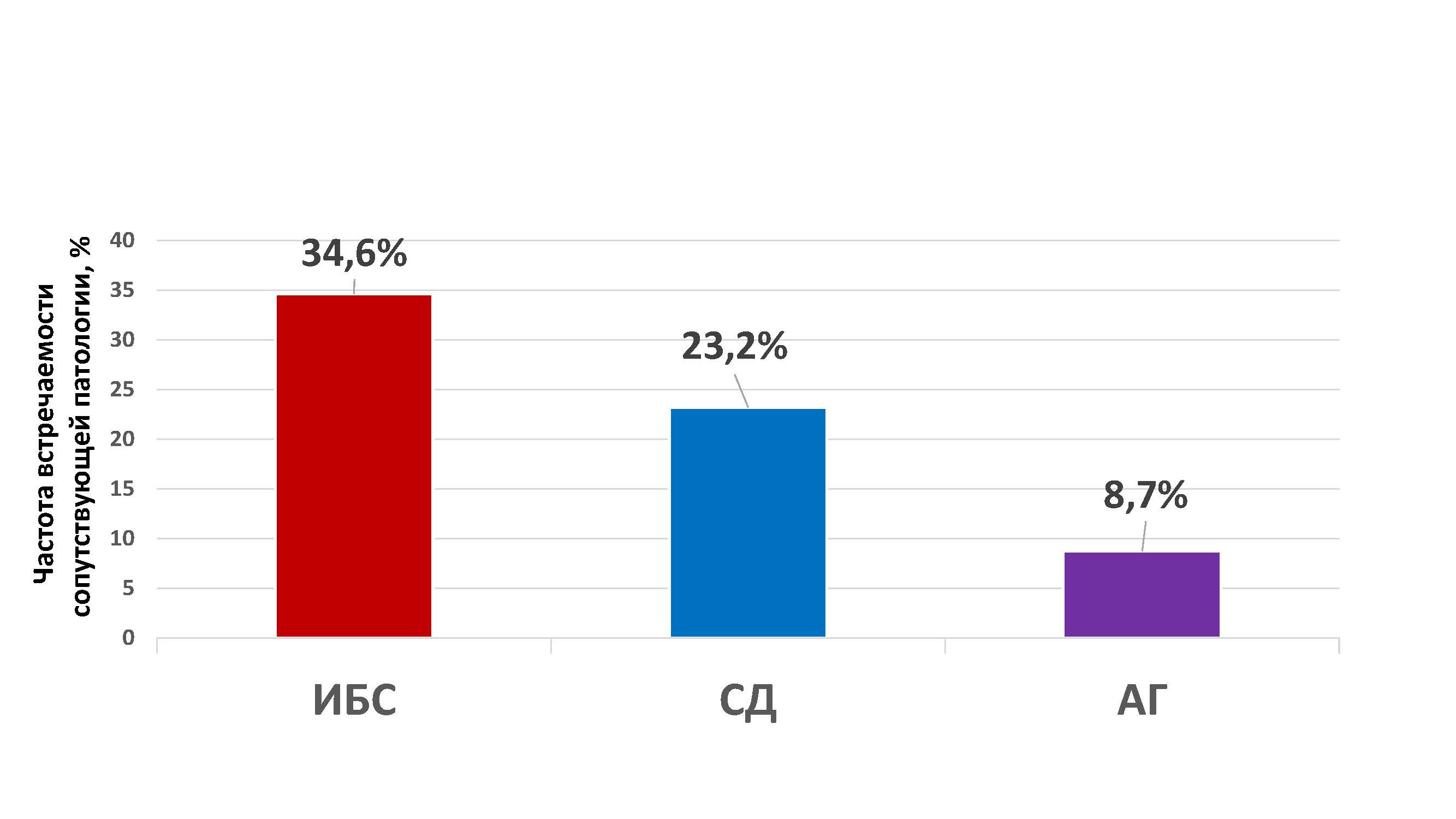
Fig. 1. Schematic representation of the incidence of comorbidities in the general group of stroke patients (n=492)
Note: CHD — coronary heart disease; DM — type 2 diabetes mellitus; AH — arterial hypertension.
According to instrumental studies, pathology in the form of visualization of morphological changes was detected on CT — in 2.6% (n=10 out of 383), on MRI — in 2.0% (n=7 out of 342), and on ultrasound — in 28.0% (n=124 out of 443) of examined patients for each instrumental modality.
The results of genetic studies in the overall patient group (n=492) are presented in Table 2.
Table 2. Genetic characteristics of patients in the general group (n=492)
| Indicator | General group (n=492) |
|---|---|
| C677T, CC, n (%) | 200 (40.7) |
| C677T, CT, n (%) | 209 (42.5) |
| C677T, TT, n (%) | 83 (16.9) |
| A1298C, CC, n (%) | 279 (56.7) |
| A1298C, CT, n (%) | 155 (31.5) |
| A1298C, TT, n (%) | 58 (11.8) |
| MTR, CC, n (%) | 163 (33.1) |
| MTR, CT, n (%) | 197 (40) |
| MTR, TT, n (%) | 132 (26.8) |
| MTRR, CC, n (%) | 199 (40.4) |
| MTRR, CT, n (%) | 210 (42.7) |
| MTRR, TT, n (%) | 83 (16.9) |
| AGT174, CC, n (%) | 393 (79.9) |
| AGT174, CT, n (%) | 77 (15.7) |
| AGT174, TT, n (%) | 22 (4.5) |
| AGT235, CC, n (%) | 316 (64.2) |
| AGT235, CT, n (%) | 88 (17.9) |
| AGT235, TT, n (%) | 88 (17.9) |
| NOS3, CC, n (%) | 306 (62.2) |
| NOS3, CT, n (%) | 83 (16.9) |
| NOS3, TT, n (%) | 103 (20.9) |
| AGTR, CC, n (%) | 377 (76.6) |
| AGTR, CT, n (%) | 68 (13.8) |
| AGTR, TT, n (%) | 47 (9.6) |
Subsequently, a comparison of the studied parameters was performed in two groups: patients with stroke (n=426) and patients with transient ischemic attack (n=66), the results of which are presented in Table 3.
Table 3. Clinical, demographic, laboratory and instrumental characteristics of patients when compared in two groups: stroke (n=426) and TIA (n=66)
| Indicator | Stroke group (n=426) | TIA group (n=66) | p |
|---|---|---|---|
| Age, Me (Q1-Q3), years | 73 (64-82) | 66 (49-79) | 0.005* |
| Male sex, n (%) | 185 (43.4) | 18 (27.3) | 0.013* |
| Female sex, n (%) | 241 (56.6) | 48 (72.7) | |
| Height, Me (Q1-Q3), cm | 168 (162-175) | 165 (160-172) | 0.048* |
| Body weight, Me (Q1-Q3), kg | 80 (67-91) | 75 (65-88) | 0.07 |
| BMI, Me (Q1-Q3), kg/m² | 27.1 (24.5-30.8) | 26.6 (24.2-30.8) | 0.468 |
| Glucose, Me (Q1-Q3), mmol/L | 6.6 (5.6-8.1) | 6.3 (5.7-7.3) | 0.197 |
| Cholesterol, Me (Q1-Q3), mmol/L | 5.1 (4.1-6.5) | 5.4 (4.6-6) | 0.678 |
| HDL, Me (Q1-Q3), mmol/L | 1.7 (1.3-2.1) | 1.6 (1.4-1.9) | 0.326 |
| LDL, Me (Q1-Q3), mmol/L | 3.1 (2.2-4.1) | 3.5 (2.9-4.1) | 0.07 |
| Atherogenic coefficient, Me (Q1-Q3) | 1.9 (1.4-2.7) | 2 (1.7-2.7) | 0.160 |
| TG, Me (Q1-Q3), mmol/L | 1.2 (0.9-1.7) | 0.93 (0.7-1.4) | <0.001* |
| K⁺, Me (Q1-Q3), mmol/L | 4.6 (4.2-4.9) | 4.2 (4.1-4.4) | <0.001* |
| Na⁺, Me (Q1-Q3), mmol/L | 142 (140-145) | 143 (139-146) | 0.782 |
| TSH, Me (Q1-Q3), mIU/L | 2.1 (1.3-2.8) | 2 (1.7-2.5) | 0.638 |
| Leukocytes, Me (Q1-Q3), 10⁹/L | 7.3 (6.1-8.9) | 7.8 (6.9-9.9) | 0.041* |
| Lymphocytes, Me (Q1-Q3), 10⁹/L | 1.6 (1.2-2.1) | 1.5 (1.1-2) | 0.173 |
| IG, Me (Q1-Q3) | 0.3 (0.2-0.6) | 0.25 (0.1-0.5) | 0.072 |
| Platelets, Me (Q1-Q3), 10⁹/L | 229 (183-275) | 233 (197-264) | 0.744 |
| Hemoglobin, Me (Q1-Q3), g/L | 138 (126-147) | 134 (128-144) | 0.470 |
| Hematocrit, Me (Q1-Q3), % | 40.1 (36.9-42.8) | 39.2 (37.5-42.3) | 0.358 |
| CHD, n (%) | 150 (35.2) | 20 (30.3) | 0.435 |
| DM, n (%) | 102 (23.9) | 12 (18.2) | 0.302 |
| AH, n (%) | 36 (8.5) | 7 (10.6) | 0.638 |
| Pathology on CT, n (%) (n=383) | 10 (3.1) | 0 (0) | 0.377 |
| Pathology on MRI, n (%) (n=443) | 124 (32.9) | 0 (0) | <0.001* |
| Length of hospitalization, days, Me (Q1-Q3) | 11 (8-14) | 10 (6-12) | <0.001* |
Note: * — differences are statistically significant (p < 0.05).
It was established that patients with stroke were statistically significantly older compared to TIA patients (p=0.005). Among them, males were more common than females (p=0.013). When comparing anthropometric data, a trend toward greater height and body weight was noted in stroke patients; however, differences in body mass index did not reach statistical significance.
In biochemical blood analysis, patients with ischemic stroke had significantly higher triglyceride levels (p < 0.001) and potassium concentration (p < 0.001) compared to TIA. However, leukocyte levels were higher in the TIA group than in the stroke group (p=0.041).
Furthermore, pathological changes on MRI were more frequently detected in stroke patients (p < 0.001). Length of hospitalization was also greater in the stroke group (p < 0.001).
The distribution of genotype frequencies of the studied polymorphic variants with the results of comparison with Hardy-Weinberg equilibrium is presented in Table 4.
Table 4. Distribution of genotypes of the studied polymorphic variants of genes with calculation of compliance with Hardy-Weinberg equilibrium in the studied patients (n=492)
| Polymorphism | Genotype | n | % | Chi-square | p |
|---|---|---|---|---|---|
| C677T | CC | 200 | 40.7 | 4.869 | 0.057 |
| CT | 209 | 42.5 | |||
| TT | 83 | 16.9 | |||
| A1298C | CC | 279 | 56.7 | 21.832 | <0.001* |
| CT | 155 | 31.5 | |||
| TT | 58 | 11.8 | |||
| MTR | CC | 163 | 33.1 | 18.89 | <0.001* |
| CT | 197 | 40 | |||
| TT | 132 | 26.8 | |||
| MTRR | CC | 199 | 40.4 | 4.54 | 0.053 |
| CT | 210 | 42.7 | |||
| TT | 83 | 16.9 | |||
| PAI | CC | 133 | 27.0 | 2.633 | 0.104 |
| CT | 228 | 46.3 | |||
| TT | 131 | 26.6 | |||
| AGT174 | CC | 393 | 79.9 | 37.05 | <0.001* |
| CT | 77 | 15.7 | |||
| TT | 22 | 4.5 | |||
| AGT235 | CC | 316 | 64.2 | 145.84 | <0.001* |
| CT | 88 | 17.9 | |||
| TT | 88 | 17.9 | |||
| NOS3 | CC | 306 | 62.2 | 173.23 | <0.001* |
| CT | 83 | 16.9 | |||
| TT | 103 | 20.9 | |||
| AGTR | CC | 377 | 76.6 | 121.78 | <0.001* |
| CT | 68 | 13.8 | |||
| TT | 47 | 9.6 |
Note: * — statistically significant deviation of genotype distribution from Hardy-Weinberg equilibrium at p < 0.05.
When testing for compliance with Hardy-Weinberg equilibrium, a statistically significant deviation from expected distribution was found for the following polymorphisms: A1298C (p < 0.001), MTR (p < 0.001), AGT174 (p < 0.001), AGT235 (p < 0.001), NOS3 (p < 0.001), and AGTR (p < 0.001). Thus, the polymorphisms whose distribution did not show a statistically significant deviation from Hardy-Weinberg equilibrium were: C677T (0.057), PAI (p=0.104), and MTRR (0.053).
Comparative genetic data in two groups: patients with stroke (n=426) and patients with TIA (n=66) are presented in Table 5.
Table 5. Comparative data of genetic studies in two groups: patients with stroke (n=426) and patients with TIA (n=66)
| Indicator | Stroke group (n=426) | TIA group (n=66) | p |
|---|---|---|---|
| C677T, CC, n (%) | 186 (43.7) | 14 (21.2) | <0.001* |
| P₁=0.001 | |||
| P₃<0.001 | |||
| C677T, CT, n (%) | 179 (42) | 30 (45.5) | |
| C677T, TT, n (%) | 61 (14.3) | 22 (33.3) | |
| A1298C, CC, n (%) | 245 (57.5) | 34 (51.5) | 0.386 |
| A1298C, CT, n (%) | 134 (31.5) | 21 (31.8) | |
| A1298C, TT, n (%) | 47 (11) | 11 (16.7) | |
| MTR, CC, n (%) | 136 (31.9) | 27 (40.9) | 0.074 |
| MTR, CT, n (%) | 179 (42) | 18 (27.3) | |
| MTR, TT, n (%) | 111 (26.1) | 21 (31.8) | |
| MTRR, CC, n (%) | 167 (39.2) | 32 (48.5) | 0.356 |
| MTRR, CT, n (%) | 186 (43.7) | 24 (36.4) | |
| MTRR, TT, n (%) | 73 (17.1) | 10 (15.2) | |
| PAI, CC, n (%) | 117 (27.5) | 16 (24.2) | 0.663 |
| PAI, CT, n (%) | 194 (45.5) | 34 (51.5) | |
| PAI, TT, n (%) | 115 (27) | 16 (24.2) | |
| AGT174, CC, n (%) | 349 (81.9) | 44 (66.7) | 0.011* |
| P₁=0.004 | |||
| P₂=0.039 | |||
| AGT174, CT, n (%) | 61 (14.3) | 16 (24.2) | |
| AGT174, TT, n (%) | 16 (3.8) | 6 (9.1) | |
| AGT235, CC, n (%) | 289 (67.8) | 27 (40.9) | <0.001* |
| P₁<0.001 | |||
| P₃<0.001 | |||
| AGT235, CT, n (%) | 72 (16.9) | 16 (24.2) | |
| AGT235, TT, n (%) | 65 (15.3) | 23 (34.8) | |
| NOS3, CC, n (%) | 273 (64.1) | 33 (50) | 0.031* |
| P₁=0.028 | |||
| P₃=0.008 | |||
| NOS3, CT, n (%) | 72 (16.9) | 11 (16.7) | |
| NOS3, TT, n (%) | 81 (19) | 22 (33.3) | |
| AGTR, CC, n (%) | 327 (76.8) | 50 (75.8) | 0.952 |
| AGTR, CT, n (%) | 59 (13.8) | 9 (13.6) | |
| AGTR, TT, n (%) | 40 (9.4) | 7 (10.6) |
Note: * — differences are statistically significant (p < 0.05).
Table 6. Calculation of sample power for comparing the distribution of genotypes between groups of patients with stroke and TIA
| Polymorphism | χ² 2×3 | p | Effect size (V Cramer's/w Cohen's) | Power, % | N for 80% | N for 90% |
|---|---|---|---|---|---|---|
| C677T | 19.4909 | <0.001 | 0.1990 | 98.3 | 243 | 319 |
| A1298C | 1.9034 | 0.386 | 0.0622 | 21.6 | 2490 | 3271 |
| MTR | 5.2028 | 0.074 | 0.1028 | 52.1 | 911 | 1197 |
| MTRR | 2.0641 | 0.356 | 0.0648 | 23.2 | 2297 | 3016 |
| PAI | 0.8224 | 0.663 | 0.0409 | 11.7 | 5764 | 7570 |
| AGT174 | 8.8987 | 0.011 | 0.1345 | 76.6 | 533 | 700 |
| AGT235 | 20.4368 | <0.001 | 0.2038 | 98.7 | 232 | 305 |
| NOS3 | 7.4216 | 0.024 | 0.1228 | 68.3 | 639 | 839 |
| AGTR | 0.0979 | 0.952 | 0.0141 | 5.7 | 48421 | 63595 |
Note: Power is calculated post-hoc for the actual observed effect size with α=0.05 and df=2. The N values for 80% and 90% reflect the estimated total sample size required to achieve the appropriate power while maintaining the observed effect size.
When analyzing genetic markers in the two comparison groups (Table 5), statistically significant differences were found with predominance in the stroke patient group for the following polymorphisms:
C677T of the MTHFR gene (CC and TT genotypes comparison: for CC p=0.001, for TT p < 0.001);
AGT174 (p=0.004 and p=0.039);
AGT235 (p < 0.001);
NOS3 (p=0.028 and p=0.008).
No significant differences between the groups were found for the remaining studied polymorphisms.
According to the literature, genetic polymorphisms affecting folate cycle enzymes (MTHFR: 677 C>T, 1298 A>C; MTR: 2756 A>G; MTRR: 66 A>G) are associated with an increased risk of cerebrovascular events, including stroke [11, 12]. Single nucleotide substitutions in the coding sequences of these genes provoke dysfunction of the corresponding enzymes, leading to disruption of folate metabolism. The consequence of enzymatic deficiency is hyperhomocysteinemia — a pathological increase in plasma homocysteine concentration. Clinical studies demonstrate that homocysteine levels have greater prognostic value for cardiovascular pathologies than total cholesterol levels. Hyperhomocysteinemia correlates with stenotic lesions of major arteries; deep vein thrombosis; microangiopathic changes; cerebrovascular events (especially in patients with coronary heart disease, chronic kidney disease, type 2 diabetes mellitus). In individuals with venous thrombosis, hyperhomocysteinemia often serves as the only identifiable risk factor. Experimental data indicate that an increase in homocysteine concentration of 20-30% above reference values can increase the risk of irreversible ischemic changes. An increase in homocysteine levels of 5 μmol/L above the upper limit of normal is associated with a 60% increase in the risk of atherosclerosis in men and 80% in women [8].
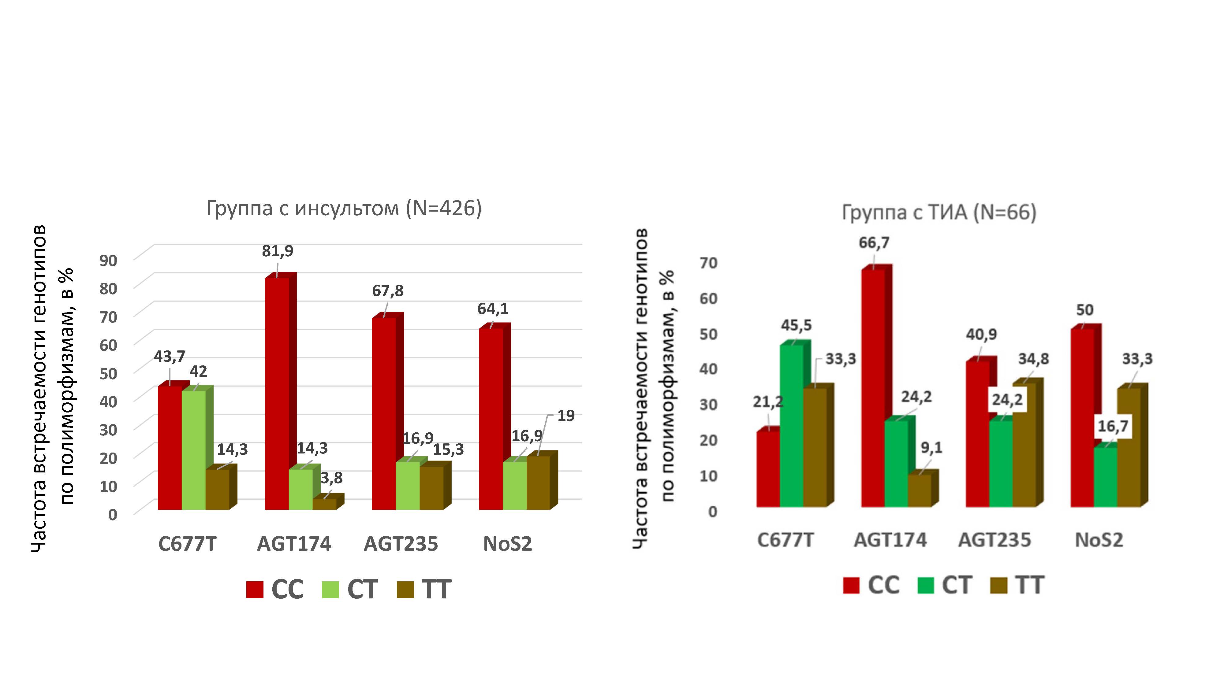
Fig. 2. Comparison of the genotype prevalence rates for the following polymorphisms: C677T of the MTHFR gene, AGT174, AGT235, and NOS3 in patients with stroke (n=426) and patients with transient ischemic attack (n=66)
Note: Only statistically significant results (p < 0.05) according to the data in Table 4 are presented.
Disorders in the functioning of the renin-angiotensin system (RAS) are associated with an increased risk of cerebrovascular events. Imbalance in RAS function can initiate the development of arterial hypertension, atherosclerotic changes, and other pathological processes that provoke cerebral lesions [13].
The AGT gene, located in chromosomal region 1q42.2, encodes the precursor protein angiotensinogen — a key substrate in the cascade of angiotensin I formation. Among the identified polymorphic variants, the rs699 (p.Met235Thr) variant has particular clinical significance, in which methionine is replaced by threonine at position 235 of the polypeptide chain. This mutation is associated with a 10-20% increase in serum angiotensinogen concentration, which correlates with an increased risk of developing arterial hypertension and cerebrovascular events, including stroke [14, 15].
Also well studied and associated with adverse risks is the AGT gene polymorphism rs4762 (p.Thr174Met), which is a point mutation resulting in the replacement of threonine (T) with methionine (M) at position 174 of the angiotensinogen protein. This structural change may have a regulatory effect on the rate of angiotensinogen proteolysis, thereby influencing the dynamics of angiotensin I generation [13].
On chromosome 7 at position 7q36.1 is the NOS3 gene, whose product — endothelial NO synthase — is an enzyme responsible for the biosynthesis of nitric oxide (NO). This process is fundamentally important for the regulation of vascular tone, blood flow, and a number of other physiological functions [14].
Despite the total number of patients (N=492) presented in the study, which is sufficient to obtain statistically reliable data for most clinical-laboratory and instrumental studies, the results of genetic testing for individual genetic polymorphic variants require a larger number of observations (due to insufficient sample power) for the evidence base necessary for future implementation of the obtained data into clinical practice.
This study confirmed the presence of a complex of clinical-laboratory and genetic differences between patients with ischemic stroke and TIA. The complex of differences identified in patients with ischemic stroke — including older age, male predominance, elevated anthropometric and biochemical parameters (triglycerides, potassium) against the background of leukopenia, prolonged hospitalization, and signs of severe brain vascular damage on MRI — fully aligns with the spectrum of known risk factors, which confirms the results of the comparative analysis with patients with transient ischemic attack.
The key result was the identification of specific genetic patterns associated with different forms of acute cerebrovascular accidents. In stroke patients, homozygous CC genotypes predominated for key polymorphisms regulating homocysteine metabolism (C677T of the MTHFR gene), angiotensinogen (AGT174, AGT235), and nitric oxide synthase (NOS3). In contrast, patients with transient ischemic attack (TIA) more frequently had alternative allelic variants: TT genotypes for C677T of the MTHFR gene, AGT235, and NOS3 polymorphisms, as well as the heterozygous CT genotype for AGT174. The identified differences in genotype distribution reflect the different genetic basis of the studied pathological conditions and can be used as additional molecular markers to improve differential diagnosis between stroke and TIA in routine clinical practice.
Thus, molecular genetic diagnostics are becoming an essential tool in the specialist's arsenal. It provides an objective basis for assessing individual genetic risk of developing persistent cerebrovascular pathology, and in the future — for developing personalized treatment algorithms taking into account pathogenetic mechanisms and targeted prevention of thrombotic complications.
1. Dutova T. I., Banin I. N., Ermolenko N. A. Genetic passport as a basis for primary and secondary prevention of cerebral infarction in young people. Lechaschi Vrach. 2023; 7-8 (26): 45-51. (In Russ.).
2. Dichgans M, Beaufort N, Debette S, Anderson CD. Stroke Genetics: Turning Discoveries into Clinical Applications. Stroke. 2021 Aug;52(9):2974- 2982. doi: 10.1161/STROKEAHA.121.032616.
3. Chugunova S.A., Nikolaeva T.Ya., Ignatieva A.V., Popov M.M. Genetic risk factors of thrombofilia in a young ischemic stroke patient (clinical case report). Yakut Medical Journal. 2017;(3):65-68. (In Russ.).
4. Temprano-Sagrera G, Sitlani CM, Bone WP, et al. Multi-phenotype analyses of hemostatic traits with cardiovascular events reveal novel genetic associations. J Thromb Haemost. 2022 Jun;20(6):1331-1349. doi: 10.1111/jth.15698.
5. Drozdova EL, Komkova GV, Polonikova AA, et al. Relationship between polymorphism rs1546155 of the GGT7 gene and the risk of ischemic stroke. Research Results in Biomedicine. 2024;10(3):339- 350. (In Russ.).
6. Tsalta-Mladenov M, Levkova M, Andonova S. Factor V Leiden, Factor II, Protein C, Protein S, and Antithrombin and Ischemic Strokes in Young Adults: A Meta-Analysis. Genes (Basel). 2022 Nov 9;13(11):2081. doi: 10.3390/genes13112081.
7. Shibeeb S, Al-Rayashi N, Shams N, et al. Factor V Leiden (R506Q), Prothrombin G20210A, and MTHFR C677T Variants and Thrombophilia in Qatar Biobank Participants: A Case Control Study. Pathophysiology. 2024 Oct 21;31(4):608- 620. doi: 10.3390/pathophysiology31040044.
8. Ni J, Zhang L, Zhou T, et al. Association between the MTHFR C677T polymorphism, blood folate and vitamin B12 deficiency, and elevated serum total homocysteine in healthy individuals in Yunnan Province, China. J Chin Med Assoc. 2017 Mar;80(3):147-153. doi: 10.1016/j.jcma.2016.07.005.
9. Huang X, Ye Q, Zhang Y, et al. Effects of eNOS rs1799983 and DYRK1A rs720470 on Susceptibility to Ischemic Stroke and its Subtypes: A Prospective Observational Study. Clin Appl Thromb Hemost. 2025 Jan-Dec;31:10760296251384918. doi: 10.1177/10760296251384918.
10. Clinical guidelines: Ischemic stroke and transient ischemic attack. https://cr.minzdrav.gov.ru/previewcr/814_1 Russian
11. Meng H, Huang S, Yang Y, et al. Association Between MTHFR Polymorphisms and the Risk of Essential Hypertension: An Updated Meta-analysis. Front Genet. 2021 Nov 26;12:698 590. doi: 10.3389/fgene.2021.698590.
12. Chang G, Kuai Z, Wang J, et al. The association of MTHFR C677T variant with increased risk of ischemic stroke in the elderly population: a meta-analysis of observational studies. BMC Geriatr. 2019 Nov 27;19(1):331. doi: 10.1186/s12877-019-1304-y.
13. Isordia-Salas I, Santiago-Germán D, Cerda-Mancillas MC, et al. Gene polymorphisms of angiotensin-converting enzyme and angiotensinogen and risk of idiopathic ischemic stroke. Gene. 2019 Mar 10;688:163-170. doi: 10.1016/j.gene.2018.11.080.
14. Jalel A, Midani F, Fredj SH, et al. Association of BglII Polymorphism in ITGA2 and (894G/T and -786T/C) Polymorphisms in eNOS Gene With Stroke Susceptibility in Tunisian Patients α2 Gene Polymorphism in α2β1 Integrin and eNOS Gene Variants and Stroke. Biol Res Nurs. 2021 Jul;23(3):408-417. doi: 10.1177/1099800420977685.
15. Kumar A, Sharma R, Misra S, et al. Relationship between methylenetetrahydrofolate reductase (MTHFR) gene (A1298C) polymorphism with the risk of stroke: A systematic review and meta-analysis. Neurol Res. 2020 Nov;42(11):913- 922. doi: 10.1080/01616412.2020.1798107.
Natalia A. Kovtun — Cand. Sci. (Med.), Head of Laboratory at Clinical Hospital No. 1 of the Presidential Administration of the Russian Federation, and Associate Professor of the Department of Healthcare Organization and Public Health, Medical Insurance, and State Control in Healthcare at the Central State Medical Academy of the Presidential Administration of the Russian Federation
Moscow
Tatyana V. Isaeva — Cand. Sci. (Med.), Head of the Medical Rehabilitation Center
Moscow
Marina I. Savelyeva — Dr. Sci. (Med.), Professor, Professor of the Department of Therapy named after Professor E. N. Dormidontov
Yaroslavl
Valery V. Boyarintsev — Dr. Sci. (Med.), Professor, Professor of the Russian Academy of Sciences, Head of the Department of Emergency Care, Urgent and Extreme Medicine
Moscow
Kovtun N.A., Isaeva Т.V., Savelyeva M.I., Boyarintsev V.V. The role of single nucleotide polymorphisms in predicting individual stroke risk. Pharmacogenetics and Pharmacogenomics. 2026;(2):24-34. (In Russ.) https://doi.org/10.37489/2588-0527-0008. EDN: BQRZEP
НАШИ КНИГИ




Другие журналы
"Издательства ОКИ"




ПАРТНЕРЫ


 Email this article (Login required)
Email the author (Login required)
Email this article (Login required)
Email the author (Login required)  Post a Comment (Login required)
121357, Moscow, st. Vatutina, 4, building 2, apt. 22
LLC "Izdatelstvo OKI"
Tel. + 7 (916) 986-04-65
Email: eva88@list.ru